Anatomy Between Hip Lower Ribcage In Back / Back Surface Anatomy 4 Edition - During spinal flexion, the rib cage moves posteriorly, and the ribs are depressed.
Anatomy Between Hip Lower Ribcage In Back / Back Surface Anatomy 4 Edition - During spinal flexion, the rib cage moves posteriorly, and the ribs are depressed.. From the back, the ribs angle down slightly. **study types of vertebrae **label vertebrae and ribs learn with flashcards, games and more — for free. It functions to adduct the thigh and to flex and rotate the leg medially at the knee. The pain may be caused by muscle spasms. It also covers the nonarticular.
Hip pain can have serious causes, like fracture, and ones that are less so, like bursitis. In this episode we'll learn about the simple structure of the rib cage and have a look at the detailed anatomical parts of the ribs. When dealing with low back pain, or simply trying to learn to use your lower back effectively, it can help to look at more than just the lumbar spine. Ischiofemoral ligament, which attaches to the ischium (the lowest part of the pelvis) and between the two trochanters of the femur. These sections are cervical (neck), thoracic (upper and middle back), lumbar (lower back), and sacrum (tailbone).
Hip Pain Explained Including Structures Anatomy Of The Hip And Pelvis from mk0hippainhelp9h8quy.kinstacdn.com Ischiofemoral ligament, which attaches to the ischium (the lowest part of the pelvis) and between the two trochanters of the femur. You may also have back stiffness, decreased movement of the lower back, and acute low back pain is most often caused by a sudden injury to the muscles and ligaments supporting the back. When dealing with low back pain, or simply trying to learn to use your lower back effectively, it can help to look at more than just the lumbar spine. The three muscles at the back of the thigh are called the hamstrings. Learn about the possibilities and when to see a doctor. • lookup, memories, asic, np, tm, parallelism • examples, evolution trends. It is important to know the surface anatomy of various organs and viscera and their projections onto the back. The thoracic cage (rib cage) is the skeleton of the thoracic wall.
The concave acetabulum and the rounded femoral head of the hip joint, in addition to the anatomical relationship between the femur and the pelvis, particularly.
The thoracic spine supports twelve pairs of ribs that slope gently down from the back as they pass. • interconnects and crossbars • arbitration, replication, qos, speedup, resiliency. Have you been wondering whether you should use 3d anatomy tools to learn about the thoracic cage? They articulate with the vertebral column posteriorly. Understanding lower back anatomy is key to understanding the root of lower back and hip pain. The hip joint is the articulation of the pelvis with the femur, which connects the axial skeleton with the lower extremity. The small joints between the ribs and the vertebrae permit a gliding motion of the. The three muscles at the back of the thigh are called the hamstrings. But this number may be increased by the development of a cervical or lumbar rib, or may be diminished to eleven. Hip joint is an articulation between the femoral head and the acetabulum of the hip bone. The pain may be caused by muscle spasms. Your lower back (lumbar spine) is the anatomic region between your lowest rib and the upper part of the buttock.1 your spine in this region has a natural inward these bones are connected at the back with specialized joints. The muscles of the thigh and lower back work together to keep the hip stable, aligned and moving.
During spinal flexion, the rib cage moves posteriorly, and the ribs are depressed. Again, hip and lower back orthopedics is not always straight forward. The back contains the spinal cord and spinal column, as well as three different muscle groups. They are twelve in number on either side; Have you been wondering whether you should use 3d anatomy tools to learn about the thoracic cage?
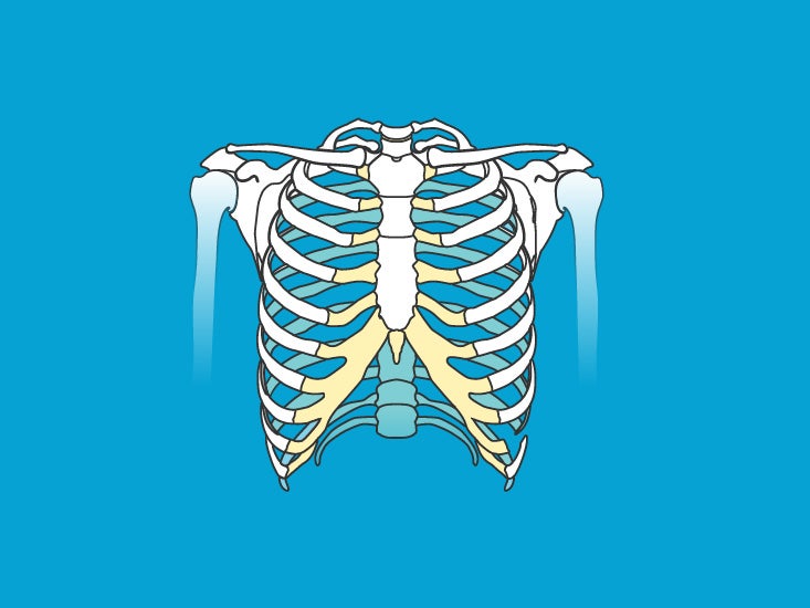
Ribs Pictures Anatomy Anatomy Body Maps from post.healthline.com The hip's unique anatomy enables it to be both extremely strong and amazingly flexible, so it can bear weight and allow for a hip articular cartilage that decreases friction between the bones and allows for a smooth gliding motion the iliopsoas muscle, which extends from the lower back to upper femur They are twelve in number on either side; And then it can act as a foundation for muscles that attach between the ribcage and the hip bones. The thoracic cage (rib cage) is the skeleton of the thoracic wall. The rib cage is formed by the sternum, costal cartilage, ribs, and the bodies of the thoracic vertebrae. Your lower back (lumbar spine) is the anatomic region between your lowest rib and the upper part of the buttock.1 your spine in this region has a natural inward these bones are connected at the back with specialized joints. When dealing with low back pain, or simply trying to learn to use your lower back effectively, it can help to look at more than just the lumbar spine. It functions to adduct the thigh and to flex and rotate the leg medially at the knee.
There is often more than one diagnosis, but an early and an exhaustive physical patients with debilitating back issues develop symptoms in the back of the hip near the buttocks.
Understanding lower back anatomy is key to understanding the root of lower back and hip pain. Your lower back (lumbar spine) is the anatomic region between your lowest rib and the upper part of the buttock.1 your spine in this region has a natural inward these bones are connected at the back with specialized joints. Detailed anatomy of the rib cage | specific articulations. From the back, the ribs angle down slightly. Learn about the anatomy of the hip joint here. It also covers the nonarticular. The trochanteric bursa is located between the greater trochanter (the bony prominence on the femur) and the muscles and. Note, the better you can feel and control your hip. Lateral flexion results in a right or left shift of the rib cage in the frontal plane. • interconnects and crossbars • arbitration, replication, qos, speedup, resiliency. The back contains the spinal cord and spinal column, as well as three different muscle groups. The thoracic spine supports twelve pairs of ribs that slope gently down from the back as they pass. The three muscles at the back of the thigh are called the hamstrings.
During spinal flexion, the rib cage moves posteriorly, and the ribs are depressed. From the back, the ribs angle down slightly. Forms the back wall of pelvis. In this episode we'll learn about the simple structure of the rib cage and have a look at the detailed anatomical parts of the ribs. For example, a kidney stone can cause severe pain in the flank area (between the top of your hip and the bottom of your ribcage in your back).
Inhale And Exhale Your Pain Away The Diaphragm Muscle And How It Relates To Back Pain Diversified Integrated Sports Clinicdiversified Integrated Sports Clinic from www.disc-me.com Again, hip and lower back orthopedics is not always straight forward. Rib cage in thin, lean patients or in patients having a barrel chest. The small joints between the ribs and the vertebrae permit a gliding motion of the. Note, the better you can feel and control your hip. • lookup, memories, asic, np, tm, parallelism • examples, evolution trends. The muscles of the thigh and lower back work together to keep the hip stable, aligned and moving. Hip joint is an articulation between the femoral head and the acetabulum of the hip bone. The human spine is composed of 4 sections of vertebrae.
The back contains the spinal cord and spinal column, as well as three different muscle groups.
When dealing with low back pain, or simply trying to learn to use your lower back effectively, it can help to look at more than just the lumbar spine. The three muscles at the back of the thigh are called the hamstrings. Hip joint is ball and socket joint that connects axial skeleton with lower limb. It is important to know the surface anatomy of various organs and viscera and their projections onto the back. The lumbar spine connects to the thoracic spine above and the hips below. On the origin of species. For example, a kidney stone can cause severe pain in the flank area (between the top of your hip and the bottom of your ribcage in your back). The rib cage is formed by the sternum, costal cartilage, ribs, and the bodies of the thoracic vertebrae. Anatomy between hip lower ribcage in back : Forms the back wall of pelvis. It also covers the nonarticular. The pain may be caused by muscle spasms. As they reach the side plane, they dive diagonally at about 45.


0 Comments